
Figure 10.
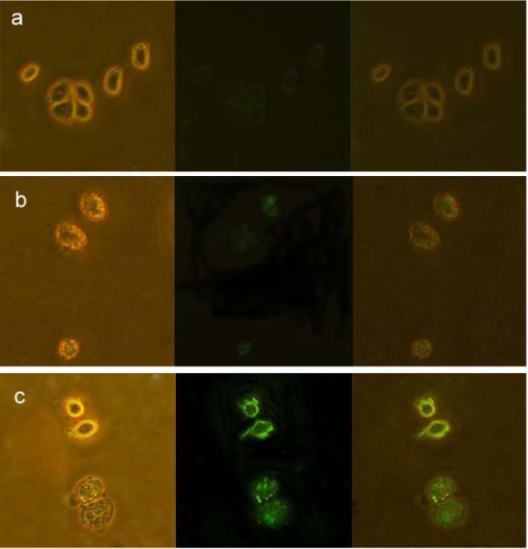
Images of live Hela cells (a) without interacting with any nanoparticles, (b) directly labeled by Fe3O4/CdTe nanocomposites without antibody conjugated and (c) immuno-labeled by Fe3O4/CdTe nanocomposites conjugated with anti-CEAcam8. In the three panels, the left rows represent the phase-contract images, the central rows represent the fluorescent images, the right rows are the overlays of the left and central rows.