Fig. 3.
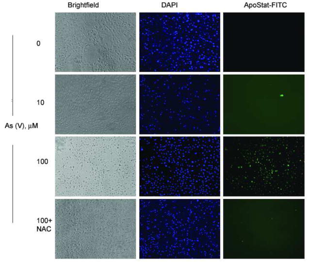
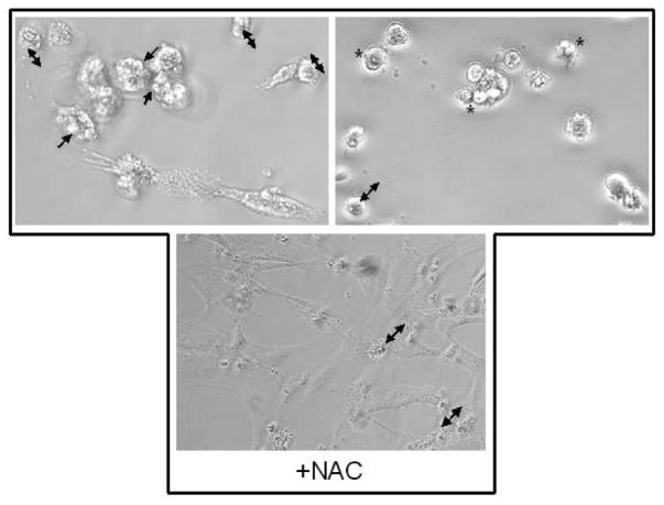
(A) Apostat ™ labeling of MEMM cells undergoing As (V) mediated apoptosis. Apostat, a FITC-conjugated, membrane permeable dye, was utilized for visualization of apoptotic cells. MEMM cells were exposed to different concentrations of As (V) for 48 hrs. Apostat was added 15 minutes before the end of the incubation period; cells were washed and co-labeled with the nuclear stain DAPI before being examined for FITC and DAPI using a Nikon TE 2000 inverted microscope equipped with FITC and UV filters. Apoptotic cells are green in the field. The photographs are representative of three identical, separate experiments. (B) Morphological assessment of MEMM cell death following treatment with 100 μM As (V). Cells were examined for membrane blebbing (→), cell condensation (↔) and formation of apoptotic bodies (*) after 48 h of treatment. Lower panel shows the effect of pretreatment with NAC. Representative photographs from three separate experiments with similar results are shown.


