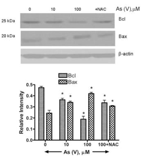
Fig. 8.

Western blot analysis of Bax and Bcl proteins in MEMM cells exposed to As (V). Cells were exposed to different concentrations of As (V) for 48 h, harvested, and lysed in a modified RIPA buffer (see Methods). One set of cells was pretreated with NAC prior to treatment with 100 μM As (V). 40 μg of total protein lysates were separated by SDS-PAGE, transferred to a PVDF membrane and immunoblotted for Bax and Bcl proteins. (A) Bax, a proapoptotic protein, has a molecular weight of 25 kDa while the antiapoptotic Bcl has a molecular weight of 20 kDa. While the levels of Bcl decrease sharply after 48 hours of 100 μM As (V) treatment, Bax levels show an increase. A change in ratio of Bcl and Bax in favor of Bax is indicative of cellular apoptosis. (B) Densitometric analysis of Western blots from three different experiments. For each treatment condition, the relative intensity of Bcl and Bax protein band was calculated by dividing its numerical intensity by that of corresponding beta-actin band.
