Figure 3.
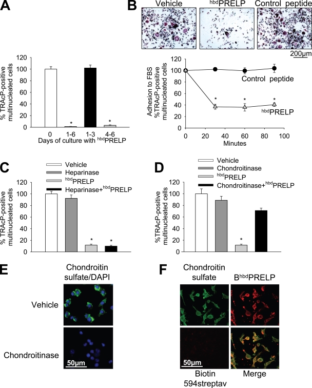
Role of cell surface proteoglycans in hbdPRELP-induced inhibition of osteoclastogenesis. (A) Purified mouse bone marrow macrophages, incubated with M-CSF and RANKL, were treated with vehicle (0) or 15 µM hbdPRELP for the entire time frame of the cultures (1–6 d), for the first 3 d (1–3), or for the last 3 d (4–6). (B) Osteoclast cultures, differentiated by M-CSF and RANKL, were trypsinized. Cells were then pretreated in suspension with vehicle, 15 µM hbdPRELP, or 15 µM of control peptide and allowed to adhere to substrate for the times indicated. Attached cells were stained for TRAcP (top) and enumerated (bottom). (C and D) Prefusion osteoclasts were pretreated with 2 U/ml heparinase III (C) or 0.45 U/ml chondroitinase ABC (D) before the addition of hbdPRELP, and then multinucleated osteoclasts were stained for TRAcP and enumerated. (E) Prefusion osteoclasts were pretreated with vehicle or with 0.45 U/ml chondroitinase ABC, fixed, and incubated with an anti–chondroitin sulfate antibody (green), with no permeabilization to assess the content of cell surface chondroitin sulfate. Cells were also stained with DAPI (blue) to detect nuclei. Pictures are the results of the merge between the two fluorescences. (F) Prefusion osteoclasts were treated with a biotin-tagged hbdPRELP (BhbdPRELP), fixed, permeabilized, and incubated with an anti–chondroitin sulfate antibody to detect colocalization of tagged hbdPRELP with cell surface and vesicular chondroitin sulfate (biotin 594-streptavidin [streptav] was used as a negative control). (A–D) Results are representative or the mean ± SEM of three independent experiments (*, P < 0.001).

