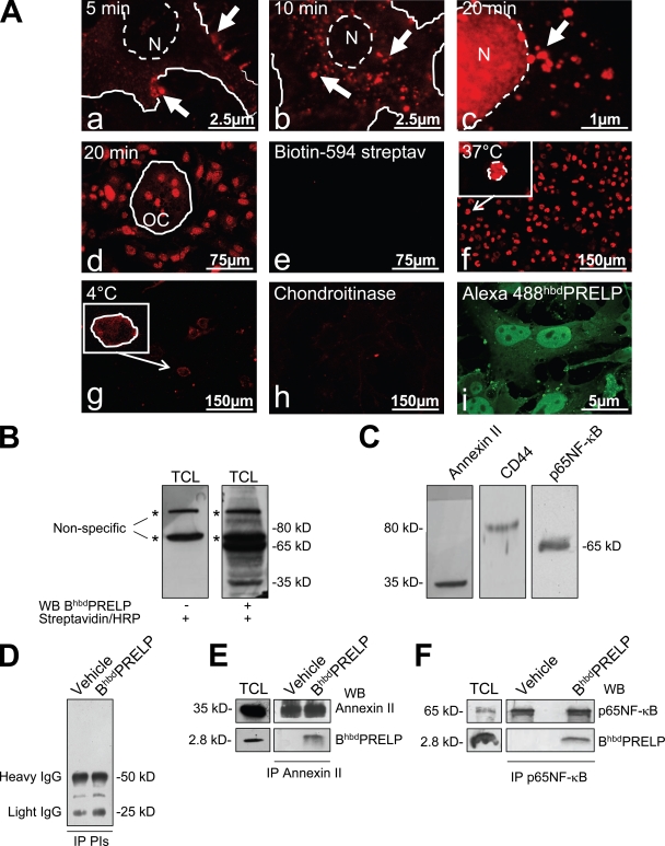
Figure 4.
Internalization of hbdPRELP and interacting proteins. (A) Vital incubation of prefusion (a–c) or mature mouse osteoclasts (OC; d) with BiotinhbdPRELP for the minutes indicated. Arrows indicate plasma membrane localization (a), localization in endosomal vesicles (b), and localization in vesicles in the vicinity of the nucleus (c). N, nuclei. (e) Negative control in the absence BiotinhbdPRELP. (f–h) Prefusion osteoclasts were incubated with BiotinhbdPRELP for 20 min at 37°C (f) or at 4°C (g) or were pretreated with 0.45 U/ml chondroitinase ABC (h) before the incubation with BiotinhbdPRELP. (f and g) Insets show higher magnification of the framed fields. (i) Fixed and permeabilized prefusion osteoclasts were incubated with Alexa Fluor 488–hbdPRELP. The solid lines indicate the cell surface, and the dashed lines indicate the nuclear envelope. (B) A prefusion osteoclast lysate was subjected to SDS-PAGE and processed with BiotinhbdPRELP (BhbdPRELP) as described in Materials and methods. Asterisks indicate nonspecific bands. (C) The filter shown in B was stripped and Western blotted for annexin II, CD44, and p65NF-κB. (D–F) Immunoprecipitation (IP) of BiotinhbdPRELP-treated prefusion osteoclast lysates with preimmune serum (D), an annexin II antibody (E), or a p65NF-κB antibody (F). Results are representative of three independent experiments. PIs, preimmune serum; TCL, total cell lysate; WB, Western blot.

