Figure 7.
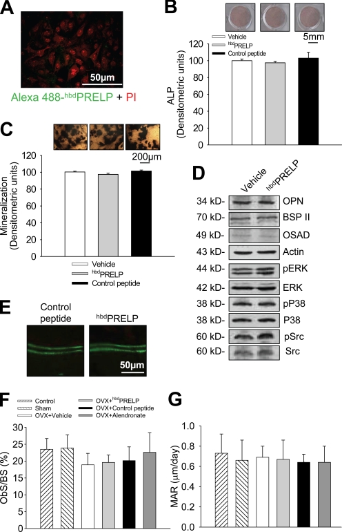
Effect of hbdPRELP on osteoblasts. (A) Calvarial osteoblasts were fixed and incubated for 1 h with Alexa Fluor 488–hbdPRELP and propidium iodide (PI). (B) Calvarial osteoblasts were incubated for 4 d with vehicle, 15 µM hbdPRELP, or 15 µM of control peptide. Cells were fixed, and ALP was detected (top) and quantified (bottom). (C) Details of von Kossa staining of mineralized nodules in calvarial osteoblasts treated with vehicle, 15 µM hbdPRELP, or 15 µM of control peptide (top) and relative quantification of mineralization in the whole cultures (bottom). (D) Western blot analysis of calvarial osteoblast lysates for the proteins indicated. (A–D) Data are representative of the mean ± SEM of three independent experiments. ERK, extracellular signal-regulated kinase; OPN, osteopontin; OSAD, osteoadherin; p, phospho. (E–G) Histomorphometric analysis of ovariectomized (OVX) mice treated with vehicle, 10 mg/kg body weight of hbdPRELP or control peptide, and 1 mg/kg body weight of alendronate, as a reference drug, 5 d/wk for 5 wk. (E) Double in vivo calcein labeling. (E–G) Data are representative or the mean ± SD of five mice per group. ObS/BS, osteoblast surface/bone surface; MAR, mineral apposition rate.

