Figure 8.
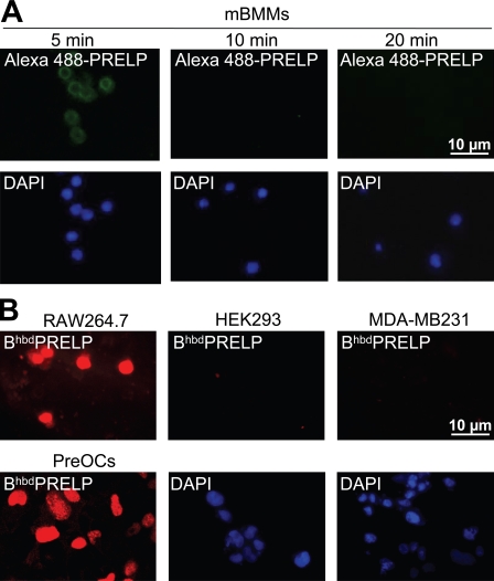
Specificity of the hbdPRELP effect. (A) Confocal microscopy of mouse bone marrow macrophages (mBMMs) vitally incubated for 5, 10, and 20 min with 15 µM Alexa Fluor 488–hbdPRELP. The bottom panels represent the nuclear DAPI staining of the cells present in the corresponding top panels. (B) Confocal microscopy of mouse osteoclast-like cells (RAW264.7), human epithelial kidney cells (HEK293), human breast cancer cells (MDA-MB231), and mouse prefusion osteoclasts (PreOCs) vitally incubated for 20 min with 15 µM BiotinhbdPRELP as described in A. The bottom middle and right panels represent the nuclear DAPI staining of the cells present in the corresponding top panels. Results are representative of three independent experiments.

