Figure 1.
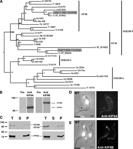
Characterization of the kinesin 9 family. (A) Phylogenetic tree constructed with kinesin 4, 6, and 9 protein sequences (348 positions), identifying two groups within the kinesin 9 family, KIF9A and KIF9B. (B) Western blot on T. brucei whole cell extracts (107 cells/lane) probed with preimmune sera (Pre), anti-KIF9A (left; 1:500) or anti-KIF9B (right; 1:500) antisera. Data were reproduced at least three times (all five mouse sera gave the same result). (C) Western blot on T. brucei cell extracts fractionated in detergent (107 cells/lane) and reproduced five times. T, total; S, supernatant (soluble fraction); P, pellet. L8C4, recognizing the PFR2 protein, was used as control. (D and E) IFA staining on WT detergent-extracted cells reproduced at least 10 times. (left) Combined phase-contrast and DAPI (white) images; (right) IFA with anti-KIF9A (D) or anti-KIF9B (E) antibodies. Values on blots are given in kilodaltons.

