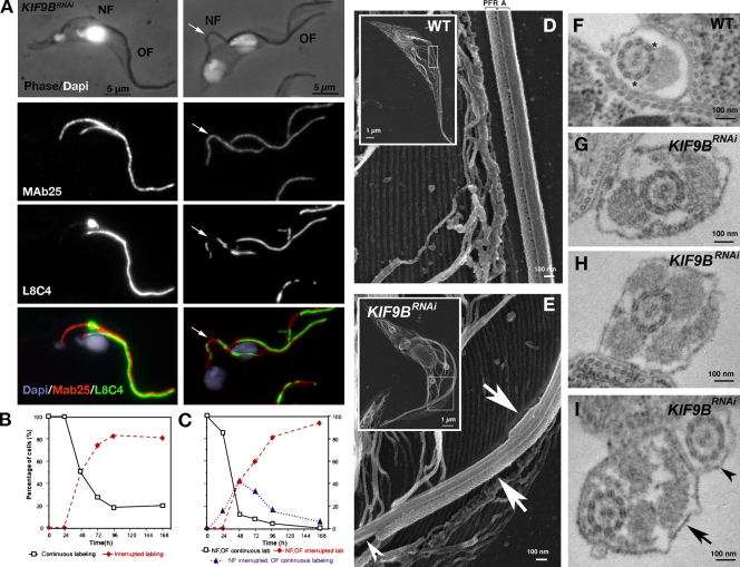
Figure 4.
Characterization of the KIF9BRNAi mutant. KIF9B is involved in PFR assembly. (A) IFA on 48 h–induced KIF9BRNAi cells. (left) Cell with one nucleus, an old flagellum (OF), and a short new flagellum (NF) are shown. The old flagellum shows a normal axoneme/PFR labeling, whereas the new flagellum shows a normal axoneme but a disrupted PFR. (right) Cells with two nuclei and two flagella exhibit the same phenotype. Detached flagella are shown by arrows. The experiment was reproduced more than three times, and at least 100 biflagellated cells were analyzed. (B and C) KIF9BRNAi cells with a normal PFR labeling (black lines) are progressively replaced by cells with an interrupted labeling (red lines) both in uniflagellated (B) and biflagellated cells (C). In biflagellated cells, the phenotype is first visible in the new flagellum (purple line). For both populations, at least 100 cells were analyzed per induction time. (D and E) Scanning electron micrograph of a WT cell (D) and a 72 h–induced KIF9BRNAi cell (E) extracted with cold Triton X-100. The white rectangle indicates the position of the magnified area. Representative images were chosen from at least 20 cells. (F–I) Transmission electron micrographs showing cross sections of flagella from WT (F) or 72 h–induced KIF9BRNAi cells (G–I). Representative images were chosen from >50 sections of WT and 89 sections of KIF9BRNAi. Asterisks indicate doublets 4–7, arrows point toward excessive PFR, and arrowheads show a naked axoneme.

