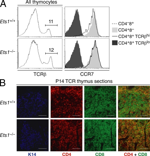
Figure 3.
Ets1−/− MHC I–restricted maturelike DP thymocytes migrate to the medulla. (A) Thymocytes from Ets1+/+ and Ets1−/− mice were stained for surface expression of CD4, CD8, TCRβ and the chemokine receptor CCR7. Overlaid histograms (right plots) analyze expression of CCR7 on Ets1−/− DP and CD4 SP thymocytes (bottom graph), and on Ets1+/+ CD8 and CD4 SP thymocytes (top graph), all TCRβhi (as gated in left plots). Expression of CCR7 on TCRlo DP thymocytes is shown in both strains as a negative control (dark gray histograms). Data are representative of two experiments. (B) Frozen thymic sections were prepared from P14 transgenic Ets1+/+ or Ets1−/− mice, stained for cytokeratin 14 (K14, pseudo-colored as blue, defining medullary areas), CD4 (red), and CD8 (green). Overlaying CD4 and CD8 staining (right) shows exclusion of DP cells from medullary areas in Ets1+/+ but not in Ets1−/− mice. The red medullary staining in Ets1+/+ mice is contributed by the few CD4 SP cells that develop in these recombination-competent animals. Bars, 100 µm. Data are representative of three experiments.

