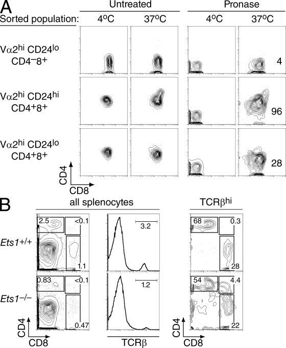
Figure 5.
Persistent CD4 expression in Ets1−/− maturelike DP thymocytes. (A) Thymocytes subsets from P14 TCR Ets1−/− mice were sorted as indicated in Fig. S3, stripped of their surface coreceptor molecules, and analyzed by flow cytometry for CD4 and CD8 surface expression after overnight single-cell suspension culture (right column). An aliquot of the pronase-treated cells was kept at 4°C and analyzed in parallel to verify the complete removal of CD4 and CD8 surface molecules after pronase digestion (third column). No change in surface coreceptor expression was seen in the absence of pronase treatment (two left columns). Data are representative of two separate experiments. Numbers in graphs indicate the mean fluorescence intensity of CD4 staining on CD8+ cells. (B) Splenocytes were prepared from 1-wk-old Ets1+/+ and Ets1−/− mice and analyzed as in Fig. 1 for expression of CD4, CD8, and TCRβ. CD4 versus CD8 two-parameter contour plots derived from TCRhi splenocytes show CD4+CD8+ splenocytes in Ets1−/− mice. Data are representative of six Ets1−/− and three Ets1+/+ neonates analyzed in two separate experiments.

