Figure 1.
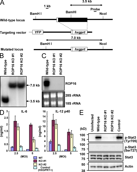
Loss of ROP16 in type I parasites severely impairs Stat3 activation. (A) The structure of the ROP16 gene, the targeting vector and the predicted disrupted gene. Black boxes denote the exons. (B) Southern blot analysis of offspring from WT or two lines of ROP16-deficient parasites. 30 µg of total genomic DNA was extracted from parasites, digested with NcoI–BamHI, electrophoresed, and hybridized with the radiolabeled probe indicated in A. Southern blotting gave a single 3.5-kb band for WT and a 7.0-kb band for the disrupted locus. (C) Northern blot analysis on 10 µg of a total parasite RNA separated on gel, transferred to a nylon membrane, and hybridized with ROP16 probe. 28S and 18S ribosomal RNA was shown as the loading control (bottom). (D) Peritoneal macrophages from C57BL/6 mice were cultured with the indicated MOI of parasites in the presence of 30 ng/ml IFN-γ for 24 h. Concentrations of IL-6 and IL-12 p40 in the culture supernatants were measured by ELISA. Indicated values are means ± SD of triplicates. (E) Serum-starved peritoneal macrophages were infected with MOI = 10 of indicated parasites for 3 h. Activation of Stat3 was also determined by Western blot of cell extracts using anti–phospho-Stat3 Tyr705 or Ser727. Stat3 and actin levels are shown as loading controls. Data are representative of at least three (D) or two (B, C, and E) independent experiments.

