Figure 2.
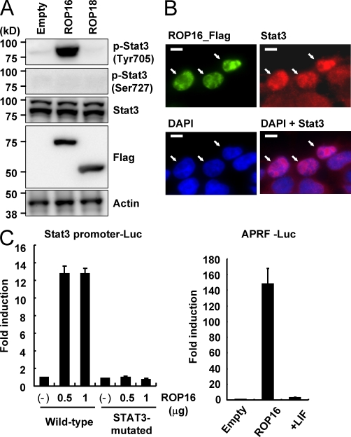
Ectopic expression of ROP16 potentiates Stat3 activation in mammalian cells. (A) 293T cells were transfected with indicated expression vectors. 48 h after transfection, the cells were lysed and subjected to Western blotting. Activation of Stat3 was also determined by Western blot analysis of cell extracts using anti–phospho-Stat3 Tyr705 or Ser727. Stat3 and actin levels are shown as loading controls. Arrows indicate cells immunostained with anti-Flag in the top left and are placed at the same positions in the other photographs. (B) 293T cells were transfected with Flag-tagged ROP16 expression vectors. 48 h after transfection, the cells were fixed and stained with anti-STAT3 or anti-Flag, and then Alexa Fluor 488–conjugated anti–mouse IgG (green), Alexa Fluor 594–conjugated anti–rabbit IgG antibody (red), or DAPI (blue). Bars, 10 µm. (C) 293T cells were transfected with indicated Stat3-dependent luciferase reporters together with either indicated amounts (left) or 1 µg (right) Flag-tagged ROP16 or empty expression vectors. As a positive control, cells were treated with 100 ng/ml human leukemia inhibitory factor (LIF) for 12 h (right). Luciferase activities were expressed as fold increases over the background level shown by lysates prepared from mock-transfected cells. Indicated values are means ± the variation range of duplicates. Data are representative of at least three (A and C) or two (B) independent experiments.

