Figure 3.
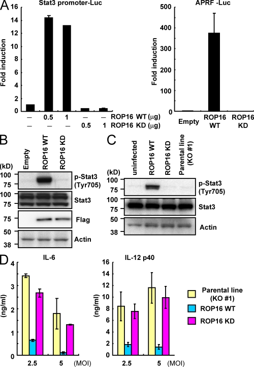
The kinase activity of ROP16 is essential for Stat3 activation. (A) 293T cells were transfected with the indicated Stat3-dependent luciferase reporters together with indicated amounts of Flag-tagged WT or kinase-inactive ROP16 (ROP16WT or ROP16KD, respectively) or empty expression plasmids. Luciferase activities were expressed as fold increases over the background levels shown by lysates prepared from mock-transfected cells. (B) 293T cells were transfected with the indicated expression vectors. 48 h after transfection, the cells were lysed and subjected to Western blotting. Activation of Stat3 was also determined by Western blot analysis of cell extracts using anti–phospho-Stat3. (C) Serum-starved MEFs were infected with an MOI = 10 of the indicated parasites for 18 h. Activation of Stat3 was also determined by Western blot analysis of cell extracts using anti–phospho-Stat3 Tyr 705. Stat3 and actin levels are shown as loading controls. (D) Peritoneal macrophages from C57BL/6 mice were cultured with the indicated MOI of parasites in presence of 30 ng/ml IFN-γ for 24 h. Concentrations of IL-6 and IL-12 p40 in the culture supernatants were measured by ELISA. Indicated values are means ± SD of triplicates. Data are representative of three (A and C), four (B), or two (D) independent experiments.

