Figure 1.
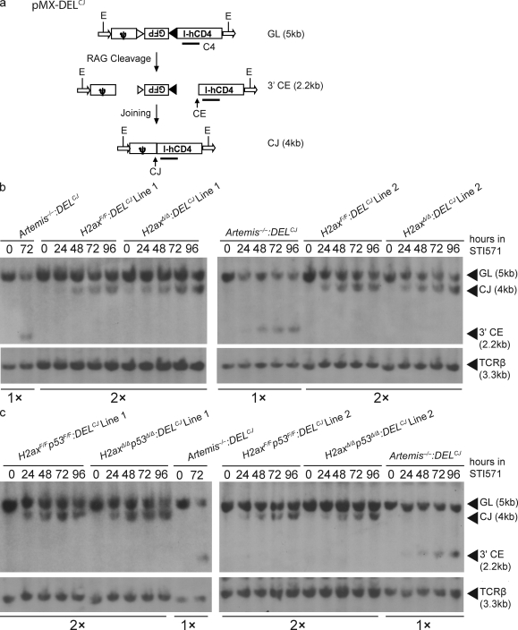
H2AX-deficient cells exhibit normal coding join formation within chromosomal substrates. (a) Shown are schematic diagrams of the pMX-DELCJ V(D)J recombination substrate in the uncleaved (GL), cleaved but not repaired (CE), and cleaved and repaired (CJ) configurations. The recombination signal sequences are represented by triangles. Arrows represent the LTR sequences. Indicated are the relative positions of the EcoRV sites (E) and C4 probe used for Southern blot analysis and the sizes of the C4-hybridizing EcoRV fragments in pMX-DELCJ substrates of the GL, CE, and CJ configuration. (b and c) Southern blot analysis of recombination products generated in cells of two different H2axF/F:DELCJ and H2axΔ/Δ:DELCJ abl pre–B cell lines (b) or H2axF/Fp53F/F:DELCJ and H2axΔ/Δp53Δ/Δ:DELCJ abl pre–B cell lines (c) treated with STI571 for the indicated times. EcoRV-digested genomic DNA was hybridized with the C4 probe. The bands corresponding to pMX-DELCJ substrates of the GL, CE, and CJ configurations are indicated. Blots were stripped and then probed with a TCR-β probe as a control for DNA content. Artemis−/−:DELCJ abl pre–B cell lines were used as a positive control for detection of pMX-DELCJ CEs, with half as much genomic DNA loaded to increase the sensitivity of detection for pMX-DELCJ CEs in experimental cells. These data are representative of experiments performed more than three independent times.

