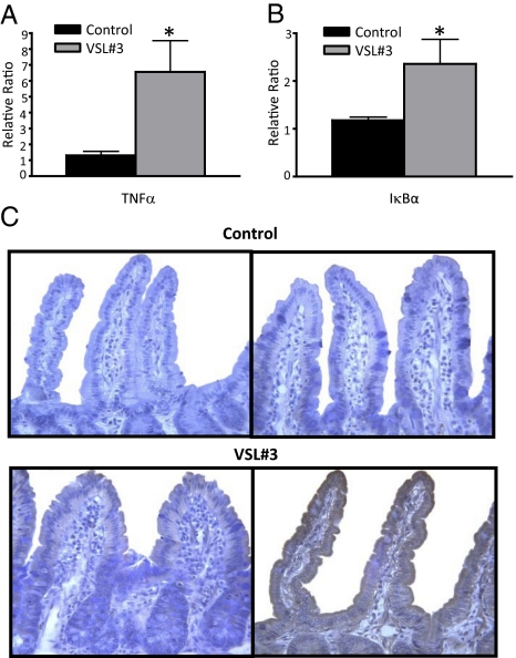
Fig. 5.
VSL#3 administration stimulates epithelial cells in vivo. (A and B) Increased expression of TNF-α and IκBα mRNA in epithelial cells after VSL#3 administration. mRNA was extracted from freshly isolated epithelial cells from SAMP mice at the end of the study period. RT-PCR was performed with specific primers and data normalized to β-actin. All values are expressed as mean ± SEM. *P < 0.05. (C) Representative photomicrographs of immunohistochemical staining demonstrated that TNF-α immunolocalized to the epithelium and was markedly increased in high-dose (Bottom Right) compared with low-dose (Bottom Left) VSL#3-treated mice, whereas untreated controls (Top Right) showed no TNF-α staining. Absence of primary detecting antibody confirmed specificity of TNF-α staining (Top Left). ×40 original magnification.

