Fig. 1.
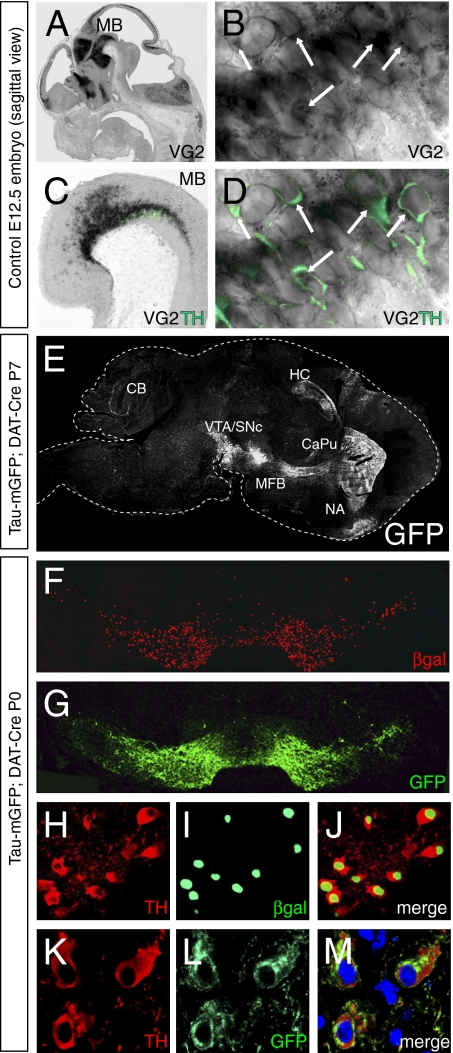
Expression of Vglut2 mRNA and DAT-Cre activity in mDA neurons. (A–D) In situ hybridization for Vglut2 mRNA combined with immunohistochemistry for TH on sagittal sections of E12.5 embryo. Vglut2 mRNA is expressed in multiple regions in the embryo, including the ventral midbrain (MB) where DA neurons develop (A). (B) Arrows indicate Vglut2-expressing cytoplasm. Vglut2 mRNA is expressed in TH-immunopositive (green) DA neurons in the MB (Magnification: B ×300) (C). (D) Arrows indicate Vglut2/TH double-positive cells (Magnification: D ×300). (E) Sagittal section of a TaumGFP;DAT-Cre brain at P7 with immunofluorescence for GFP visualizing the mDA neurons in the VTA and SNc and their projection pathway in the median forebrain bundle (MFB) to the target neurons in the striatum (CaPu + NA). GFP projections also are seen in the hippocampus (HC) and the cerebellum (CB). (F and G) Coronal sections of newborn (P0) TaumGFP;DAT-Cre mouse showing immunohistochemistry for β-gal (red) and GFP (green) in the VTA and SNc. (H–L) Coronal sections from TaumGFP;DAT-Cre P0 mouse showing TH (H, red) and β-gal (I, green) immunohistochemistry within the same cells (merged fluorescence in J), and TH (K, red) and GFP (L, green) within another set of cells (merged in M).

