Fig. 1.
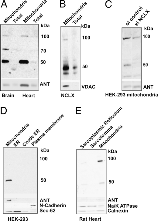
NCLX is localized to mitochondria. (A) Immunoblot analysis of NCLX in total tissue lysate (Total) and mitochondrial fractions (Mitochondria) purified from mouse heart and brain (15 μg). (B) Immunoblot analysis of total cellular and mitochondrial fractions purified from HEK-293-T cells overexpressing murine NCLX (10 μg). (Lower) Immunoblots of ANT or VDAC serving as markers. (C) Immunoblot analysis of NCLX in mitochondrial fractions purified from HEK-293 cells transfected with siNCLX or scrambled siRNA (siControl). Note that siNCLX diminishes expression levels of both the 50-kDa and 100-kDa forms of NCLX. (D and E) Expression of NCLX in cellular and tissue fractions of the indicated components purified from HEK-293 cells (20 μg) (D) or rat heart (10 μg) (E). (Lower) Immunoblots of ANT (mitochondrial marker), Na+/K+ ATPase, or N-cadherin (plasma membrane, PM, marker) and Calnexin or Sec-62 (ER, marker). Note that the mitochondria are the major site of NCLX localization (the slight NCLX signal in cardiac sarcoplasmic reticulum is presumably related to cross-contamination with mitochondria; see ANT staining).

