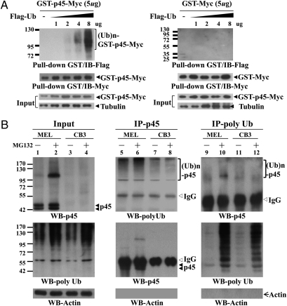
Fig. 3.
Poly-ubiquitination of p45. (A) Poly-ubquitination of p45 in transfected 293T cells. The cells were cotransfected with pEF-GST-p45-Myc or pEF-GST-Myc with different amounts of pEF-Flag-Ub. At 45 h posttransfection, the cells were treated with 10 μM MG132 for 3 h. The WCE were then prepared, and the Myc-tagged GST-fusion proteins were isolated by GST pull-down assay. Then, they were analyzed by WB with anti-Flag and anti-Myc antibodies. (B) Poly-ubiquitination of the endogenous p45 in erythroid cell lines. The MEL and CB3 cells were treated with 20 μM MG132 for 3 h, and the WCE were prepared for analysis by IP. The IP samples were then analyzed by WB with uses of anti-p45, anti-FK2 (for poly-Ub), and anti-actin antibodies.

