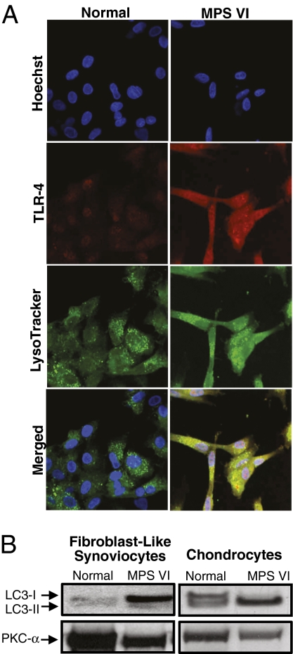
Fig. 2.
Localization of TLR4 in MPS VI lysosomes. (A) Rat FLSs were incubated with TLR4 primary polyclonal anti-rat antibody and LysoTracker Green to visualize lysosomes. Visualization of TLR4 was accomplished using a fluorescent secondary antibody, Cy-3, and nuclei were stained with a bis-benzamide Hoechst dye. Note the elevated levels of TLR4 in MPS VI cells and colocalization with the LysoTracker Green dye. (B) Anti-LC3-I (14 kDa) and LC3-II (16 kDa) Western blots of FLSs and articular chondrocytes from 6-month-old normal and MPS VI rats showing the lack of conversion of the LC3-I isoform to LC3-II in the FLSs. In contrast, in MPS VI articular chondrocytes, LC3-I was completely converted to LC3-II. Protein kinase C-α (Pkc-α) was used as a loading control.

