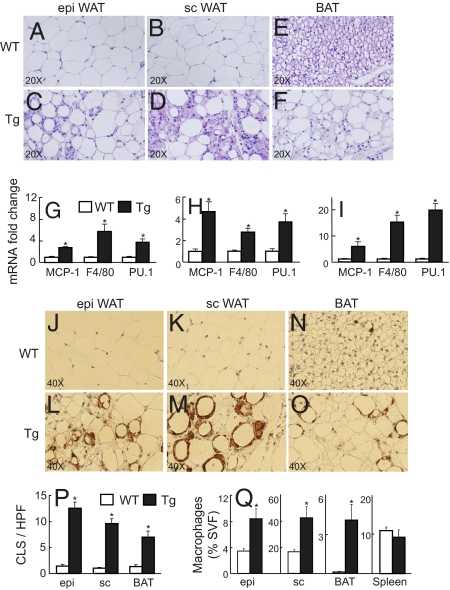
Fig. 2.
Adipose tissue histology and FACS analysis. (A–F) Histological sections from epi (A, C) and s.c. (B, D) WAT, and BAT (E, F) from representative 24-week-old WT control and Tg littermates stained with H&E. (G–I) mRNA expression from epi (G) and sc (H) WAT, and BAT (I) from WT control (open bars) vs. Tg (filled bars) littermates (n = 6). (J–O) Immunohistochemical localization of macrophages in epi (J, L), sc (K, M), and BAT (N, O) of a representative WT control vs. Tg littermates. (P) Counting of crown-like structures (CLS) per high power field (HPF) from the above male WT control and Tg littermates (10 HPF counted per mouse, n = 5 mice per genotype). (Q) Flow cytometry analyses of macrophage number from epi, sc, BAT, and spleen WT controls vs. Tg littermates. n = 4, *P < 0.05 WT vs. Tg.

