Fig. 3.
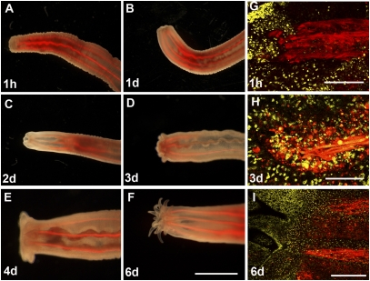
Muscle reorganization during head regeneration. (A–F) A live MyHC11::mCherry G1 transgenic polyp regenerating a head after bisection below the pharynx. The ectoderm at the wound bends inward and starts to form a new pharynx. The first tentacle buds appear after 3 days of regeneration. Note that more than 14 tentacles appear at once. (G–I) Confocal closeups of the regenerating tip (same orientation as in A–F). ToPro-3–stained nuclei are shown in yellow. (G) Retractor muscles retract immediately after being cut from the wound. (H) At day 3 after bisection, numerous cells expressing the mCherry transgene under control of the MyHC1 promoter accumulate at the wound site. (I) After 6 days, mesenteries are attached near the differentiated pharynx close to the base of the tentacles. [Scale bars: 500 μm (A–F), 20 μm (G and H), and 40 μm (I).]

