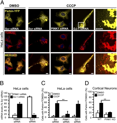
Fig. 2.
PINK1 knockdown prevents Parkin recruitment to depolarized mitochondria. (A) Immunofluorescence of HeLa cells cotransfected with Parkin-YFP and scrambled (scr) PINK1 or DJ-1 siRNA, and incubated for 1 h with 10 μM CCCP. Mitochondria are labeled with an anti-TOM20 antibody (red). (Scale bars, 10 μM.) Zoom shows 6× magnification of the region outlined by the box. (Scale bars, 1 μM.) (B) Effects of siRNA on PINK1 and DJ1 mRNA levels. Total RNA extracted from each sample is quantified by real-time PCR (n = 3). (C) Percentages of cells from the same set of cotransfected HeLa cells as in (A) that exhibit Parkin puncta colocalizing with the mitochondrial marker TOM20 (Parkin+/TOM20+). (D) Percentages of WT and knockout (KO) PINK1 cortical neurons that exhibit Parkin+/TOM20+ puncta following 1-h incubation with or without 100 nM CCCP. Values represent means ± SD (n = 30–50 cells) and are representative of 2–3 independent experiments. **, Different from controls (Newman-Keuls post hoc test; P < 0.001).

