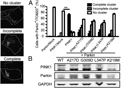
Fig. 5.
PINK1 PD mutations mitigate the formation of perinuclear mitochondrial clusters. (A) Three types of mitochondrial network morphology are defined in Parkin- and PINK1-cotransfected cells: no cluster (i.e., normal mitochondrial tubular network and distribution); incomplete cluster (i.e., mixture of perinuclear clustered mitochondria and dispersed linear mitochondria); and complete cluster (i.e., all mitochondria are clustered at the perinuclear area). Cells cotransfected with WT Parkin and PINK1 show mainly complete clusters while cells cotransfected with WT Parkin and PINK1 disease mutations or artificial dead kinase mutation (K219M) show mainly incomplete or no clusters. Bars represent percentage of cells for each type of mitochondrial morphology ± SEM; n = 200 cells counted during 3 independent experiments. **, Different from PINK1/Mock (Newman-Keuls post hoc test; P < 0.01). (Scale bars, 5 μm.) (B) Western blot from total cell extracts showing that Parkin and PINK1 expression levels were comparable in WT PINK1 and PINK1 mutants L347P and W437×. Parkin is immunodetected using anti-myc antibody. Loading is normalized with GAPDH.

