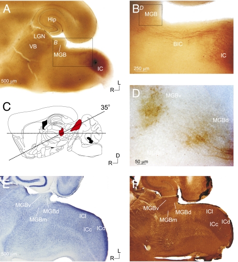
Fig. 1.
Anatomical and histological characterization of the auditory tectothalamic slice. The slice was prepared with a 35° blocking cut on the dorsal surface (C; red-shaded areas indicate the MGB and IC, respectively) [adapted from Paxinos and Franklin (14)]. Biocytin injections in the IC of the slice (A) labeled tectothalamic fibers, which traversed through the brachium of the inferior colliculus (BIC) (B) and terminated in the MGB (D). Nissl (E) and parvalbumin (F) staining delineated the IC and MGB subdivisions. (Scale bars: A, 500 μm; B, 250 μm; D, 50 μm.)

