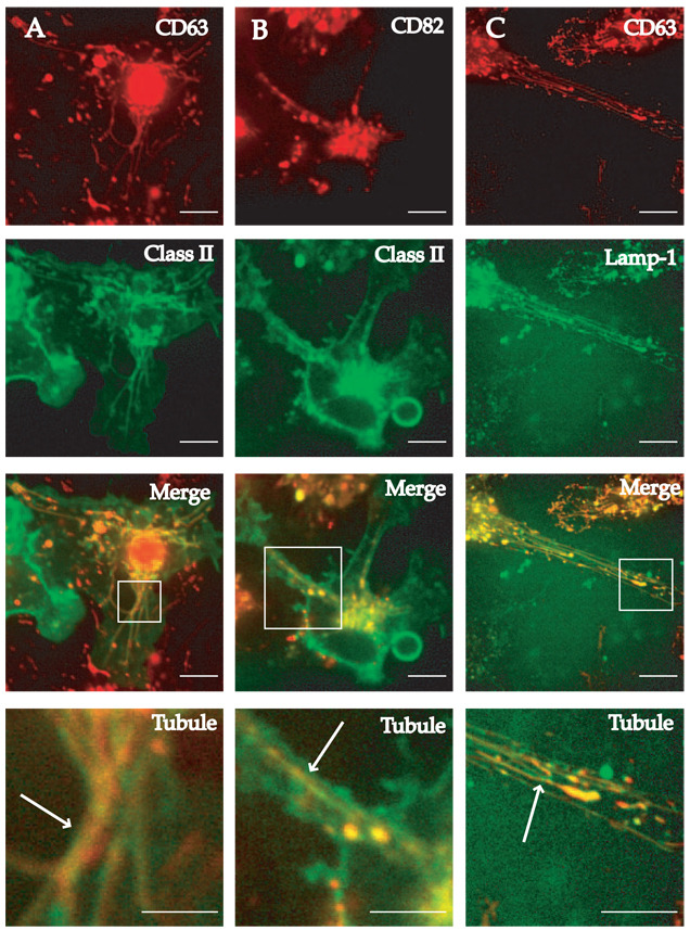
FIGURE 2.
CD63, CD82, class II MHC, and LAMP1 colocalize in endolysosomal tubules of LPS-activated BMDCs. BMDCs from class II MHC-eGFP were transduced with lentivirus encoding either CD63-mRFP1 (column A) or CD82-mRFP1 (column B) and stimulated with 100 ng/ml LPS to induce the formation of endolysosomal tubular compartments. Individual images show endolysosomal tubules composed of CD63 or CD82 (both red) and the class II compartment (green). Merged images are shown in the third row from the top. Magnification of the region of the cell demonstrating tubular compartments is shown in the bottom panel of each column. B6 BMDCs were transduced with lentiviruses expressing either CD63-mRFP1 or LAMP1-eGFP and exposed to 100 ng/ml LPS for 2 h before imaging (column C). Cells expressing both CD63 (red) and LAMP1 (green) were imaged. The merged image is shown in the third panel from the top and magnification of the tubules is seen in the bottom panel. Scale bars represent 5 µm.

