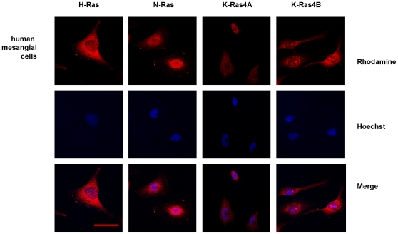
Figure 1. Immunofluorescence analysis of cellular distribution of Ras isoforms in human mesangial cells.
Pictures show H-Ras, N-Ras, K-Ras4A and K-Ras4B expression in human mesangial cells. The rhodamine red-labeled proteins are Ras isoforms, while the blue-labeled staining is Hoechst-stained DNA. Magnification: 630×. Scale bar: 30 µm. Antibodies: H-Ras, sc-520; K-Ras4A, sc-522; K-Ras4B, sc-521; N-Ras, sc-519.

