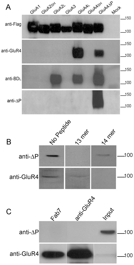
Figure 4. Analysis of AMPA receptors with an antibody specific for the exposed PDZ motif in GluA4ΔP.
(A) HEK293 cells expressing flag-tagged AMPA receptor subunits with all potential wild-type CTDs and the mutant GluA4ΔP (indicated above) were immunoblotted with the antibodies indicated to the left. Short-tailed isoforms of GluA2 and GluA4 are indicated by SH. The initial antiserum, anti-BDL detects both A2 and A4 long tails isoforms and GluA4ΔP. After the depletion procedure and purification, the anti-ΔP IgG only recognises GluA4ΔP (lower panel). (B) Anti-ΔP IgG labels a single 100 kD band in rat cerebellar tissue; this is specifically blocked by preincubation with 13mer peptide (upper panels). Similarly anti-GluR4 IgG also labels a 100 kDa band. This labelling is blocked by pre-incubation with 14 mer peptide (lower panels). (C) Immunoprecipitation from rat cerebellar extract using independent AMPA receptor antibodies fails to bring down anti-DΔP immunoreactivity (upper panel). An alternative antibody shows GluA4 levels were highly enriched in the immunoprecipitates (lower panel).

