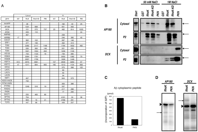
Figure 3. Identification of novel substrates for Rho-kinase.
(A) Protein scores of APP, AP180, and DCX in eluates off each affinity column. (B) Immunoblot analysis of eluates with anti-AP180 and -DCX Abs. Eluates off affinity columns with 50 mM and 1 M NaCl were subjected to immunoblot analysis with anti-AP180 and -DCX Abs. AP180 was detected in both 50 mM and 1 M NaCl eluates off the Rho-kinase-cat column, but was barely detectable in eluates off the PKN-cat column. On the contrary, DCX was strongly detected in eluates off the PKN-cat column, and moderately off the Rho-kinase-cat column. Arrows indicate the positions of AP180 and DCX. The lower bands are supposed to be degradation products or splice variants. (C) The APP cytoplasmic peptide was incubated with GST-Rho-kinase-cat or GST-PKN-cat in the presence of 100 µM [γ-32P] ATP for 1 h at 30°C. The reaction mixtures were applied to P81 paper and subjected to scintillation counting. (D) GST-AP180 (left) or GST-DCX (right) was incubated with GST-Rho-kinase-cat or GST-PKN-cat in the presence of 100 µM [γ-32P] ATP for 1 h at 30°C. The reaction mixtures were subjected to SDS-PAGE, and phosphorylated proteins were imaged by autoradiography. Arrowheads and arrows indicate substrates and autophosphorylation of Rho-kinase, respectively. These results are representatives of at least three independent experiments.

