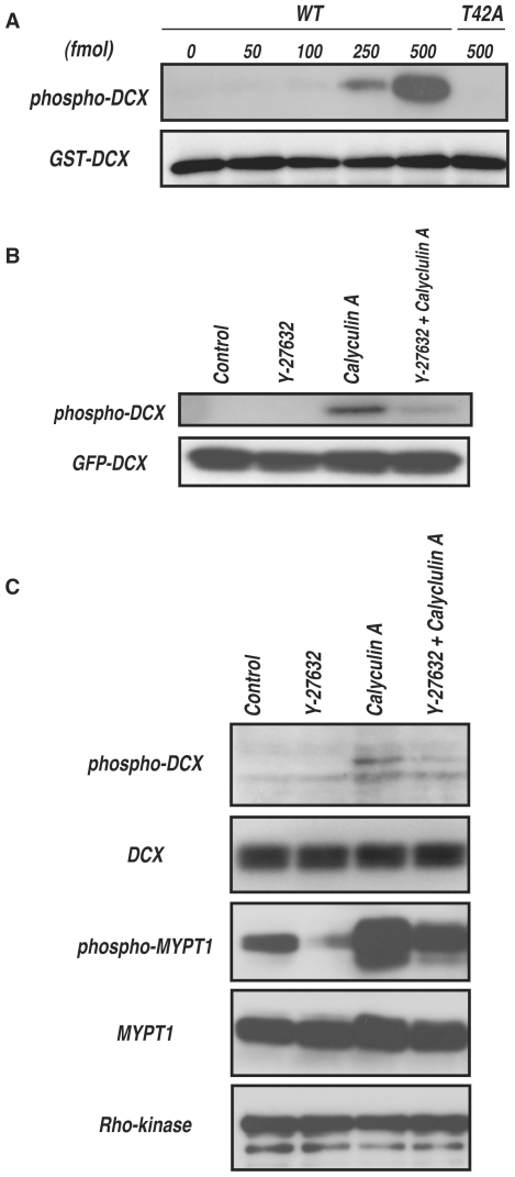
Figure 5. Phosphorylation of DCX in vivo.
(A) Specificity of the antibody against DCX phosphorylated at Thr-42 (anti-pT42 Ab). One pmol of GST-DCX containing the indicated amounts of phosphorylated GST-DCX-WT or -T42A was subjected to SDS-PAGE, followed by immunoblot analysis with anti-pT42 Ab (upper panel) or anti-GST Ab (lower panel). (B) Phosphorylation of DCX in COS7 cells. GFP-DCX was transiently expressed into COS7 cells. The transfected cells were treated with DMSO or 20 µM Y-27632 for 15 min, and then treated with or without 0.1 µM calyculin A for 10 min. The cell lysates were analyzed by immunoblot analysis with anti-pT42 Ab (upper panel) or anti-GFP Ab (lower panel). These results are representatives of at least three independent experiments. (C) Phosphorylation of DCX in hippocampal neurons at DIV1. The cells were treated with DMSO or 20 µM Y-27632 for 20 min, and then treated with or without 50 nM calyculin A for 7 min. The cell lysates were analyzed by immunoblot analysis with anti-pT42 Ab or anti-DCX Ab. Phosphorylation of MYPT1 was also examined with anti-MYPT1 pT853 Ab. These results are representatives of at least three independent experiments.

