Fig. 8.
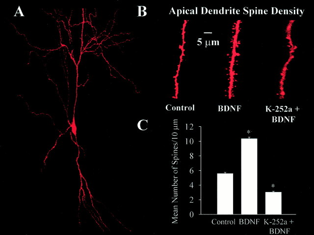
BDNF increases spine density in CA1 pyramidal neurons. A, Representative CA1 pyramidal neuron from a BDNF-treated slice filled with Alexa-594 and imaged by confocal microscopy. B, Higher magnification views of representative segments of apical dendrites from control (left), BDNF-treated (middle), and K-252a + BDNF-treated (right) slices used to quantify dendritic spine density. C, Histograms of the number of dendritic spines per 10 μm of CA1 pyramidal neuron apical dendrite. Anasterisk indicates p < 0.05.

