
Fig. 2.
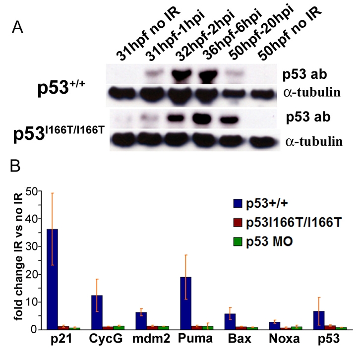
p53I166T protein has defects in transcriptional transactivation but not protein accumulation. (A) Western blots of protein extracts that were collected from WT and p53I166T/I166T mutant embryos at 1, 2, 6 and 20 hours following 0 Gy (mock IR) or 30 Gy of IR at 30 hpf. Blots were probed with p53 and α-tubulin antibodies. Note that the p53+/+ 50 hpf 20 hpi lane is underloaded. (B) RT-PCR assays for p21, cyclin G (CycG), mdm2, p53, puma, noxa and bax were performed in triplicate on WT, p53I166T/I166T and p53 MO-injected embryos. The fold induction reflects the comparison between IR-induced samples and matched non-IR samples. Error bars show the standard deviation (S.D.).