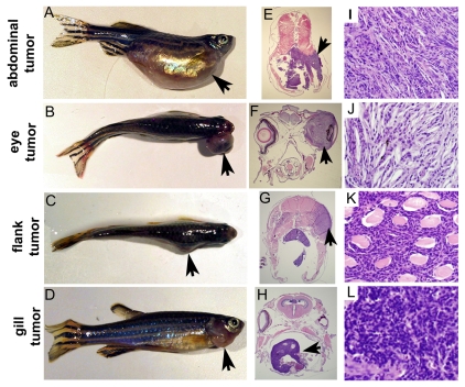
Fig. 4.
Gross and histological analysis of tumors. (A-D) p53I166T/I166T fish with abdominal (A), eye (B), flank (C) and gill (D) tumors. Sections of abdominal (E,I), eye (F,J), flank (G,K) and gill (H,L) tumors stained with hematoxylin and eosin (H&E), shown at low magnification (E-H) and high magnification (I-L). Histologically, seven of the nine abdominal tumors, all ten eye tumors, all seven flank tumors, and both gill tumors (in total, n=27/29 tumors) were spindle cell sarcomas. Arrows indicate tumors.

