
Fig. 5.
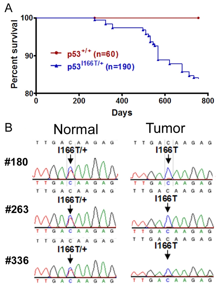
p53I166T/+ heterozygotes develop tumors and display LOH. (A) p53I166T/+ fish show a significant increase (P<0.0006) in tumor incidence compared with the p53+/+ cohorts. By 23 months, 33 of 190 p53I166T/+ fish had developed tumors. (B) Sequence analysis of the PCR products from three heterozygous fish (#180, #263 and #336) comparing normal (tail) and tumor genomic DNA. A compounded red and blue trace at codon position 166 (arrow) indicates heterozygosity, whereas a trace with blue only indicates LOH in tumor tissue samples.