Fig. 6.
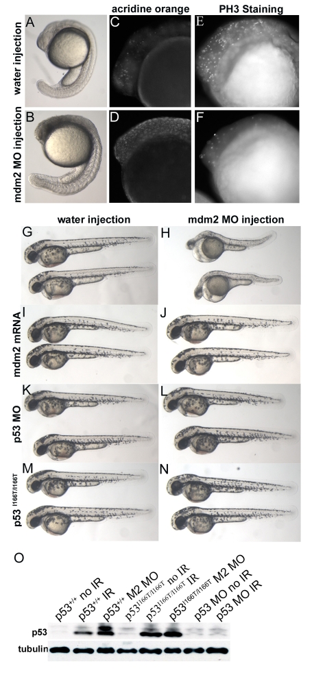
Mdm2 knockdown embryonic lethality is rescued in p53 morphants and p53I166T/I166T mutants. (A-F) Embryos injected with water (A,C,E) or Mdm2 MO (B,D,F) were assessed at 26 hpf by transmitted-light microscopy (A,B) AO staining for apoptotic cells (C,D) and PH3-specific antibody staining for cells in mitosis (E,F). Mdm2 morphants displayed embryonic lethality (B), which was probably the result of both increased apoptosis (D) and decreased cell cycling (F). With mock (water) injections as controls (G,I,K,M), Mdm2 MO injections (H) yield a readily discernable lethal phenotype at 50 hpf. (J) As a control for non-specific MO effects, co-injection of 150 pg of mdm2 mRNA rescues the Mdm2 MO phenotype. (L) Co-injection of p53 MO rescues Mdm2 MO lethality, indicating that loss of p53 function abrogates loss of the Mdm2 phenotype. (N) Injection of Mdm2 MO into p53I166T/I166T embryos did not yield the Mdm2 lethal phenotype, indicating that the p53 mutant has LOF similar to the p53 MO. (O) p53 protein levels were assessed using an anti-p53 antibody (ZFp53-9.1) on western blots of extracts from p53+/+ and p53I166T/I166T embryos that were unirradiated (no IR), irradiated with 30 Gy (IR) or injected with the Mdm2 MO (M2 MO). Extracts of wild-type embryos injected with the p53 MO are shown as controls for antibody specificity.

