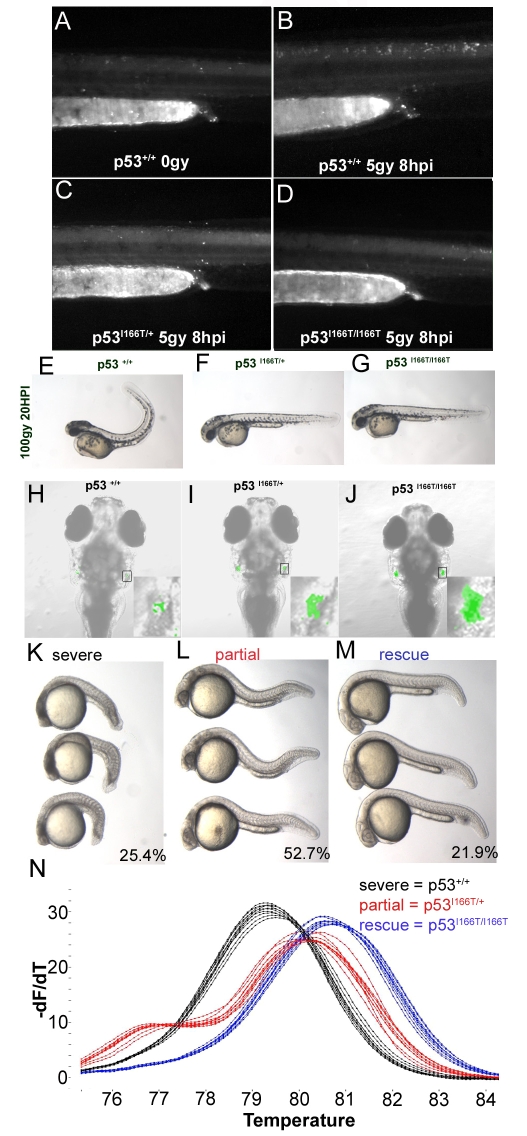
Fig. 7.
p53I166T/+ DN phenotypes. (A) The NT in the trunk and in the tail of non-irradiated p53+/+ embryos has a low level of apoptosis, detected with AO. (B) Apoptosis is increased in embryos 8 hours after they were irradiated with 5 Gy of IR. By contrast, p53I166T/+ (C) and p53I166T/I166T (D) embryos did not display increased apoptosis at 8 hours after 5 Gy of IR. (E-G) Wild-type embryos (E) have a curled tail phenotype at 20 hours after 100 Gy of IR, whereas both p53I166T/+ (F) and p53I166T/I166T (G) embryos did not display a curled tail phenotype. (H-J) Confocal analysis of EGFP expression at 9 dpf in p53+/+; Lck-EGFP/+ (H), p53I166T/+; Lck-EGFP/+ (I) and p53I166T/I166T; Lck-EGFP/+ (J) fish at 1 day after 30 Gy of IR. Insets are higher magnification views of EGFP expression. As another test for DN phenotypes, embryos from a p53I166T/+ intercross were injected with the Mdm2 MO and sorted by phenotype at 24 hpf: 25.4% had a severe lethal phenotype (K), 52.7% had a partially rescued phenotype (L) and 21.9% had a rescued phenotype (M). (N) Melting curve analysis of PCR products from individual genomic DNA samples from embryos with severe (black), partial (red) and rescued (blue) phenotypes, which correspond to the p53+/+, p53I166T/+ and p53I166T/I166T genotypes, respectively.

