Fig. 1.
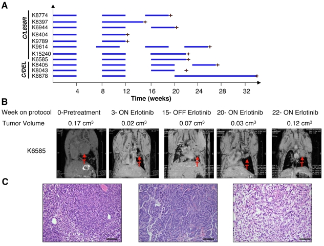
Erlotinib-resistant lung adenocarcinomas emerge after long-term intermittent drug treatment. (A) Line chart depicting the schedule used to treat individual mice with erlotinib. Mice were treated 5 days per week for 4 weeks (blue horizontal bars indicate treatment with 25 mg/kg/day of erlotinib) after which treatment was interrupted for 4 weeks (no bars). Erlotinib was administered sooner to animals that became cachectic during this period (see, for example, K9614). The treatment cycle was repeated up to three times. Doxycycline administration was initiated shortly after weaning and subsequently kept constant throughout the life of the animal. Red crosses indicate the time point at which the mouse was sacrificed. K15240, K6585 and K8043 were heterozygous for p53, and K6678 was heterozygous for Ink4a/Arf (also known as Cdkn2a). C=CC10-rtTA, L858R=TetO-EGFRL858R, DEL=TetO-EGFRΔL747-S752. (B) Coronal magnetic resonance images of lungs from a C/L858R/p53+/− (K6585) mouse subjected to the erlotinib treatment protocol. At the end of the final treatment cycle, erlotinib treatment was continued and the tumor volume increased despite the presence of the drug. Tumor volume measurements are shown. (C) Hematoxylin and eosin images of lung adenocarcinomas in mice after multiple cycles of erlotinib treatment (left and center panels). Areas of scarring where tumors had apparently regressed were also observed in the lungs of the mice (right panel). Bars, 200 μm.

