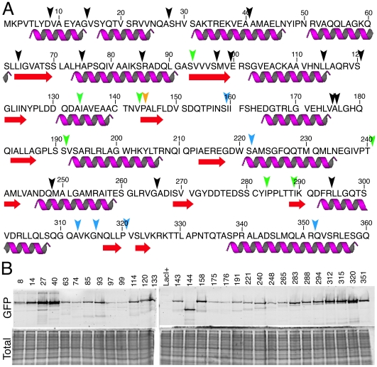
Figure 2. Analysis of TAGIT-2-constructed GFP insertions in the E. coli lactose repressor LacI.
(A) Amino acid sequence and structural features of LacI, with purple helices indicating α−helices and red arrows indicating β-sheets. Black arrowheads indicate the position of non-functional GFP insertions (Repression−, Focus−). Green arrowheads indicate GFP insertions that fail to repress lacZ, but form foci (Repression−, Focus+) when introduced into a strain with the lacO array integrated near the terminus of replication. Blue arrowheads indicate insertions that repress lacZ (Repression+, Focus+). The orange arrowhead indicates the position of the GFP insertion that is out of frame on the 3′ side of GFP. (B) The LacI-GFP insertions accumulate to variable levels. Numbers correspond to the last undisrupted lacI codon before TAGIT. Cells were harvested at an OD600 of ∼0.5, samples prepared and subject to SDS-PAGE. Protein accumulation was determined using in-gel GFP fluorescence (top panel) and the gel was subsequently stained with Coomassie blue to reveal total protein (bottom panel).

