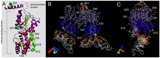
Figure 4. Cartoon of LacI-GFPi proteins mapped onto the crystal structure of LacI.
(A) The monomeric structure of LacI without the DNA binding domain (PDB ID: 1LBI). All LacI-GFPi proteins that localize as foci were mapped onto a ribbon representation of the LacI crystal structure. The amino acid corresponding to the site of GFP insertion is labeled and represented as a space-filling model. Red amino acids correspond to insertion proteins that are unable to repress the lac operon and green amino acids correspond to insertion proteins that retain some level of repression activity. (B) Model of the LacI crystal (PDB ID: 1LBG) structure including the DNA (white), DNA binding domain (orange), N-terminal core domain (blue), C-terminal core domain (light purple), and tetramerization domain (pink). The most active GFP insertion (LacI-312-GFPi) and LacI-351-GFPi are shown as green balls and two examples of inactive GFP insertions as red balls. (C) Same as in (B) but rotated to show the projection of amino acid 312 from the surface.

