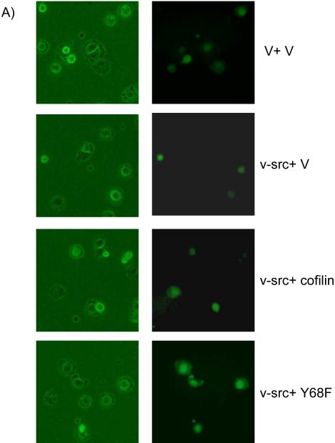
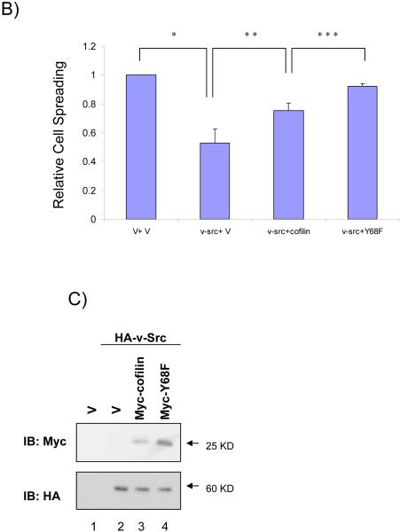
Figure 4. Reversion of v-Src inhibition of cell spreading by cofilin and Y68F mutant.
(A and B) CHO cells were transfected with Myc-tagged cofilin or Y68F mutant, and HA-tagged v-Src or vector control along with a plasmid encoding GFP as a transfection marker, as indicated. Cell spreading assays were performed as described in the Experimental Procedures. Representative micrographs are shown in A. Panel B shows the mean + S.E. of percentage of spread cells (among transfected cells as identified by GFP expression) from three independent experiments are shown as relative spreading after normalization to that in mock transfected cells. *P<0.05 in comparison with value from mock cells. **P<0.05 in comparison with value from cells transfected with v-Src and vector (v-Src + V). ***P<0.05 in comparison with value from cells transfected with v-Src and cofilin (v-Src + cofilin). (C) Aliquots of WCL were analyzed by western blotting with anti-Myc (upper) or anti-HA (lower). Molecular weight markers are indicated on the right.


