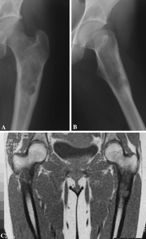
Fig. 2A–C.
A CS of the left proximal femur in seen in these radiographs. In the (A) AP and (B) lateral projections, the inner lateral and posterior cortex is invaded (moderate scalloping) by the tumor. The cortical augmentation (enlargement, Grade I) is also evident. (C) The definition of the intramedullary tumor involvement is enhanced by MRI.

