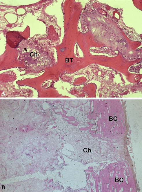
Fig. 3A–B.
The photomicrographs show the histologic presentation of Grade I CS. (A) A pattern of permeative infiltration is seen, with encasement of host bone trabeculae (BT) by the progression of the tumor (Ch) at high magnification (Stain, hematoxylin and eosin; original magnification, ×20). (B) The chondrosarcoma (Ch) invaded the host bone cortex (BC) (Stain, hematoxylin and eosin; original magnification, ×10).

