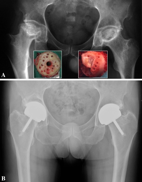
Fig. 1A–B.
(A) Anteroposterior radiograph of a 52-year-old man with bilateral steroid-induced Ficat Stage IV osteonecrosis. The insets show the extent of the femoral defects after removal of the necrotic lesions. In this case, the length of the neck could not be maintained because the femoral defects were too large to preserve a part of the chamfered area. (B) Eight years after resurfacing, the components are securely fixed and the patient’s UCLA hip scores are 10, 10, 10, and 7 for pain, walking, function, and activity, respectively.

