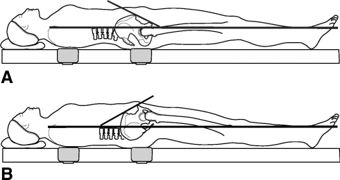
Fig. 2A–B.
(A) A diagram shows anterior pelvic tilt. The distance between the middle point of two anterior-superior iliac spines and the coronal plane is longer than that between the anterior surface of the pubic symphysis and the coronal plane. (B) Pelvic tilt is seen in this diagram. The distance between the middle point of the two anterior-superior iliac spines and the coronal plane is shorter than that between the anterior surface of the pubic symphysis and the coronal plane. (Reprinted with permission from The Association of Bone and Joint Surgeons from Wan Z, Malik A, Jaramaz B, Chao L, Dorr LD. Imaging and navigation measurement of acetabular component position in THA. Clin Orthop Relat Res. 2009;467:32–42.)

