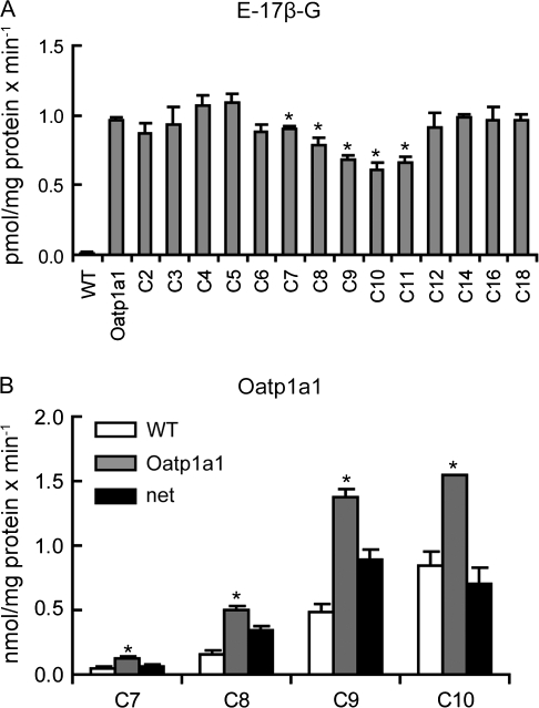
FIG. 6.
Inhibition of Oatp1a1-mediated transport and uptake of selected PFCAs by Oatp1a1. (A) Uptake of 10nM E-17β-G was measured at 37°C for 1 min with Oatp1a1-expressing or wild-type CHO cells (WT) in the absence or presence of 10μM PFCAs with chain lengths from 2 (C2) to 18 (C18) carbon atoms. Each bar represents the mean ± SD of triplicate samples *p < 0.05 compared to Oatp1a1-mediated uptake in the absence of PFCAs. (B) Uptake of 10μM C7–C10 was measured at 37°C for 1 min with wild-type (open bars) or Oatp1a1-expressing (gray bars) CHO cells. Black bars indicate net uptake after subtracting the uptake values of wild-type from Oatp1a1-expressing cells. Each bar represents the mean ± SD of triplicates. *p < 0.05 compared to vector control.

