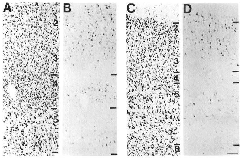
Fig. 4.
A series of brightfield photomicrographs demonstrate the laminar organization of COX 2-ir neurons in somatosensory fields of the cortex (A,C). Laminae are indicated on the adjacent Nissl-stained sections (B,D). A,B: Primary somatosensory cortex. C,D: Secondary somatosensory cortex. Scale bar = 100 μm

