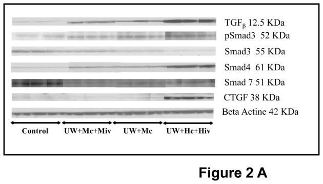
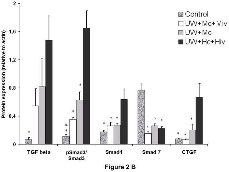
Figure 2. Western Blot analysis of the TGFβ signaling pathway in kidney graft, 3 months post transplantation.
A: representative blots for each antibody used in the four groups of survivors. B: Quantitative analysis by densitometry of blots. Psmad3 is expressed related to Smad3 expression. Shown are mean±SEM, * p < 0.05 versus UW+Hc+Hiv; ° p < 0.05 versus Control, £ p < 0.05 versus UW+Mc. n=3–5.


