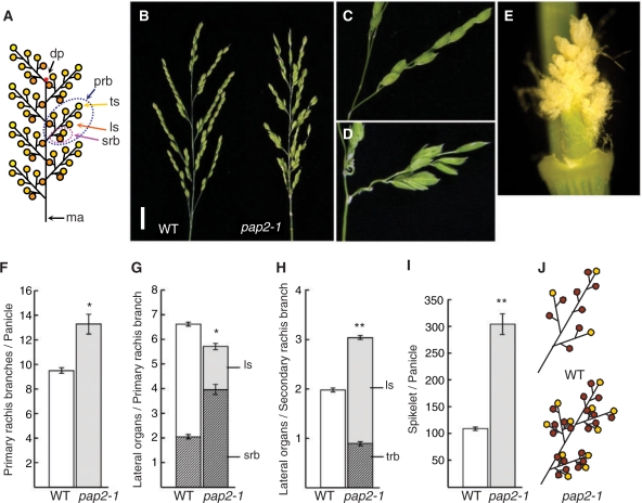
Fig. 1.
Phenotype of the pap2-1 mutant in panicle development. (A) Schematic of a rice panicle. ma, main axis; prb, primary rachis branch; srb, secondary rachis branch; ls, lateral spikelet; ts, terminal spikelet; dp, degeneration point of the IM. (B) Morphology of panicles in the wild-type (left) and pap2-1 (right). Scale bar = 2 cm. (C and D) Spikelets on the primary rachis branch in the wild-type (C) and pap2-1 (D). (E) Magnified view of aggregated branches on the pap2-1 primary rachis branch shown in D. (F) Number of primary branches per panicle. Sample size, n = 10. (G) Number of lateral organs generated on a primary branch. srb, secondary rachis branch; ls, lateral spikelet. Sample size, n = 10. (H) Number of lateral organs generated on a secondary branch. trb, tertiary rachis branch; ls, lateral spikelet. Sample size, n = 10. (I) Number of spikelets per panicle. All spikelets generated were counted under the stereomicroscope irrespective of whether they were normal or abortive. Sample size, n = 10. (J) Schematics of a spikelet of the wild-type (top) and pap2-1 mutant (bottom). The asterisks show that the difference is significant at the *1% and **0.1% level, respectively.

