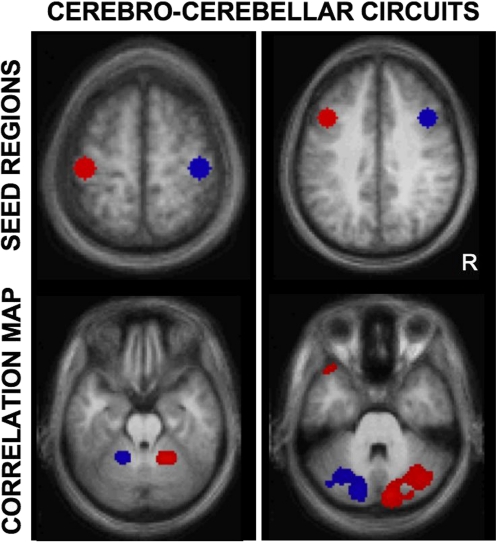
Fig. 15.
Functional connectivity reveals segregated fronto-cerebellar circuits. Two distinct cerebro-cerebellar circuits are mapped using right- and left seed regions (top) to illustrate the application of fcMRI and its sensitivity to detect anatomic circuitry. The cerebellar maps (bottom) display the functional connectivity differences between the lateralized seed regions. The maps reveal the contralateral projections of the cerebral cortex to the cerebellum and show segregated projections for motor (left) and prefrontal (right) circuits. The ability of fcMRI to map segregated, contralateral cereballar projections from the cerebral cortex demonstrates that fcMRI is constrained by anatomy and also that functional correlations reflect polysynaptic connectivity. Adapted from Krienen and Buckner (2009).

