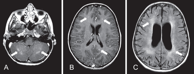
Figure 1).
Magnetic resonance imaging of the brain of a seven-year-old boy with Baylisascaris procyonis eosinophilic meningoencephailitis. A Gadolinium-enhanced T1-weighted axial image at presentation, demonstrating patchy areas of abnormal enhancement of the cerebellar grey-white junction at presentation (arrows). B Axial T1-weighted image post-gadolinium, demonstrating small foci of nodular enhancement bilaterally in the cerebral cortex at presentation (arrows). C Fluid-attenuated inversion recovery (FLAIR) axial image three months following presentation demonstrating diffuse volume loss with prominent sulci and ventricular enlargement, and white matter gliosis (arrows). The previously noted nodular enhancement had resolved

